![]()
![]()
![]()
![]()
![]()
![]()
![]()
![]()
![]()
![]()
![]()
![]()
![]()
![]()
![]()
![]()
![]()
Glaucoma in Dogs
Glaucoma is the elevation of pressure inside the eye, beyond a point at which vision is compromised or is no longer possible. Glaucoma is a frequent cause of blindness in humans and animals
There are two main types of glaucoma; Primary and Secondary.
Primary glaucoma is known to occur in certain purebred breeds of dogs and is thought to be inherited
Secondary glaucoma is caused by an external source such as a blow or a tumour.
Some breeds in which primary open-angle glaucoma is seen, are Beagles and Norwegian Elkhounds. Narrow-angle glaucoma (an abnormal narrowing of the outflow channel) is seen in American and English Cocker Spaniels. A developmental abnormality of the drainage angle called goniodysgenesis which results in decreased outflow during times of inflammation is seen in the Basset Hound, American and English Cocker Spaniel, Samoyed, and Chow Chow.
What happens?
To understand glaucoma, it is necessary to understand how the normal flow of intraocular fluid maintains normal intraocular pressure. The fluid inside the eye is called the aqueous humor .It is produced in the ciliary body which is located behind the iris. This fluid flows through the pupil and drains from the eye through a sieve-like network located at the junction of the cornea and the iris called the drainage angle. The aqueous humor is produced and drains from the eye at approximately the same rate, resulting in a stable pressure inside the eye of 15 to 30 mmHg (millimetres of mercury). Glaucoma occurs as a consequence of inadequate outflow of aqueous humor and a subsequent buildup of pressure inside the eye. The resulting high pressure damages the optic nerve and results in blindness.
Glaucoma results in blindness by blocking the nerve impulse through the optic nerve. Once the optic nerve has been permanently damaged, there can be no restoration of vision. With early aggressive and appropriate surgical intervention and then medical therapy, your pet's vision can at times be maintained. Frequently with extreme elevations of pressure, the eye becomes permanently blind and painful. The aim of therapy at that point is to keep your pet pain-free and maintain a cosmetic appearance to the eye.
DIAGNOSIS OF GLAUCOMA
The diagnosis of glaucoma is based on history, clinical signs, tonometry and gonioscopy.
Clinical signs of glaucoma include some or all of the following: excessive tearing, a green or yellow eye discharge, a reddened eye, an eye that suddenly looks blue, an eye with a pupil that is large and will not move when light is shone into it, a pet who sleeps a lot, a pet who hides under the bed or a pet who suddenly becomes frightened or irritable particularly in poor light.
In later stages of glaucoma, the eye becomes enlarged.
Tonometry is the measurement of pressure within the eye. Most veterinary ophthalmologists use the highly accurate applanation tonometer
Gonioscopy is a technique used to evaluate the drainage angle. It involves placing a goniolens - a dome-shaped contact lens - on the corneal surface after freezing the cornea with topically applied anesthetics. This lens allows the vet to directly visualize the drainage angle. Gonioscopy occasionally requires sedation but in most pets it can be performed with the use of topical anesthetics only. The technique is essential to evaluate the non-glaucomatous eye for risk of a future attack of glaucoma.
Now what happens?
TREATMENT OF GLAUCOMA
TREATMENT FOR AN EYE WITH GLAUCOMA WHERE VISION IS STILL PRESENT
Anterior Chamber Shunts
Some veterinary ophthalmologists recommend a surgical procedure where a small valve like device is implanted just under the surface of the white of the eye. This was our dog’s treatment. This device has a small tube which enters the eye through a tiny incision and this tube provides an alternate drainage pathway for the aqueous fluid to leave the eye. While some ophthalmologists report frustration with this technique since the little tube may become blocked with fibrin, or the functioning of the valve may be compromised by scarring, other ophthalmologists report considerable success with the procedure.
Other treatments include Laser surgery or freezing of the ciliary body to
reduce the amount of fluid
If your dog has become completely blind;
Another technique used to control glaucoma is the injection of gentamycin (an antibiotic) into the inside of the eye. This drug in high concentrations result in a killing effect on the ciliary body resulting in the reduction or cessation of the aqueous humor production. If the eye was visual the antibiotic injection would also kill the retina resulting in permanent blindness. Therefore, this technique can be used only on eyes that are definitely blind due to chronic pressure elevations. A brief anesthetic is required .Fluid is withdrawn and an equal amount of antibiotic is injected into the eye through the white of the eye or sclera.
Enucleation
Finally, the blind, painful eye may be surgically removed or enucleated. After enucleation, the skin is stitched shut and the hair will soon re-grow over the surgery site. This surgery again requires that your pet be anesthetized. Rarely are there any complications to this technique except possible infection.
___________________________________________________
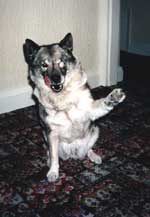
Our young dog went through all these main treatments and with the exception of her initial fear of being away from us at the Vet hospital, she coped extremely well.(Far better than we did). Eventually none of the treatments worked and the final result was the removal of one eye and total blindness.
However, she lived very happily for another 9 years and at no time did her blindness worry her. She played, ate, went on holiday with us, ran loose off lead and hunted just like any other dog. She learned to respond instantly to our commands so we could warn her about obstacles.
Here she is at around 5 years of age, after having one eye removed and the other treated with gentamycin after she had gone blind.